Jared Murray, CWC
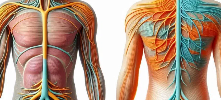
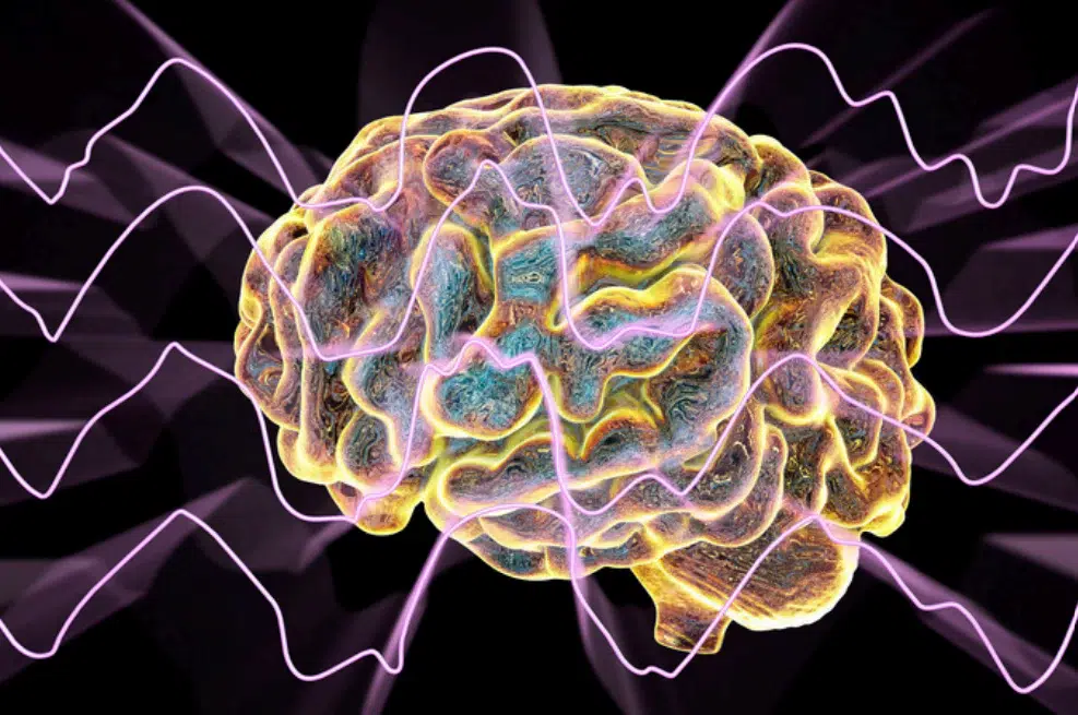
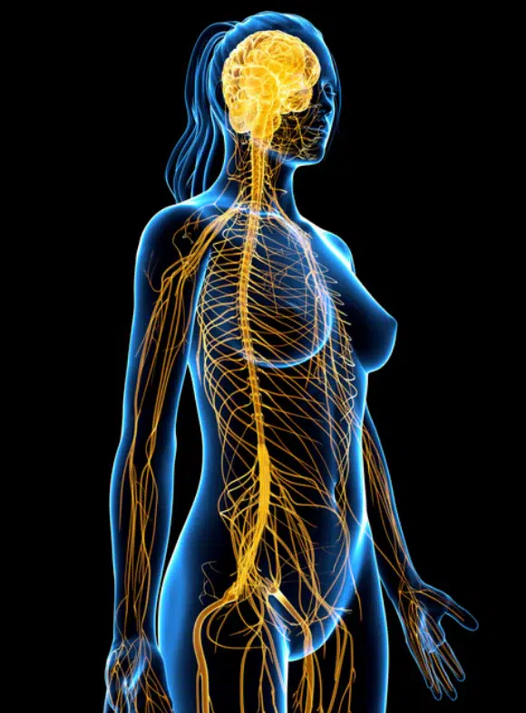
Brain wave frequencies have significant influences on various brain structures, including the hypothalamus, thalamus, amygdala, cortex, hippocampus, pituitary gland, cerebellum, Broca’s area, ventricles, and visual and auditory areas. These structures are integral to numerous physiological and psychological functions, and their activity can be modulated by different brain wave states.
The hypothalamus regulates autonomic functions and hormone production. Delta and theta waves can promote relaxation and sleep, influencing the hypothalamus to reduce stress and anxiety levels, which is beneficial for conditions like PTSD and chronic stress disorders .
The thalamus acts as a relay station for sensory and motor signals. Beta and gamma waves can enhance alertness and cognitive function, thereby improving focus and attention, which is crucial for treating ADHD and cognitive impairments.
The amygdala is involved in emotional processing. Theta waves can help in reducing fear and anxiety, aiding in the treatment of PTSD, anxiety disorders, and phobias.
The cortex is responsible for higher-order brain functions, including thought, perception, and voluntary movement. Alpha waves can promote relaxation and stress reduction, improving conditions like anxiety and depression. Beta and gamma waves enhance cognitive performance and memory, which is beneficial for learning disabilities and cognitive rehabilitation. Effects on brainwave entrainment, vagus nerve stimulation,and prenatal development.
The hippocampus is critical for memory formation and spatial navigation. Theta waves are associated with memory consolidation and learning, which can help in treating memory-related isorders and enhancing learning processes.
The pituitary gland, often called the “master gland,” regulates various hormones. Delta waves during deep sleep can facilitate hormone regulation and overall homeostasis, which is important for growth, metabolism, and stress response.
The cerebellum coordinates voluntary movements, balance, and motor control. Alpha waves can enhance coordination and reduce motor control disorders, while beta waves can improve motor learning and rehabilitation in conditions like ataxia and cerebellar disorders.
Broca’s area is involved in speech production and language processing. Beta waves can improve speech fluency and language learning, aiding in the treatment of speech and language disorders such as aphasia.
The brain’s ventricles contain cerebrospinal fluid (CSF), which cushions the brain and removes waste. Delta waves during deep sleep can promote CSF flow and brain detoxification, contributing to overall brain health and reducing the risk of neurodegenerative diseases.
The visual cortex processes visual information, while the auditory cortex processes auditory information. Gamma waves can enhance visual and auditory processing, improving conditions like dyslexia and auditory processing disorders.
Solfeggio Frequencies and Brain Structures Solfeggio frequencies, specific tones believed to have healing properties, can also influence these brain structures:
- 396 Hz: Liberating guilt and fear, beneficial for the amygdala.
- 417 Hz: Facilitating change and undoing situations, aiding the hippocampus.
- 528 Hz: Transformation and DNA repair, enhancing overall brain health.
- 639 Hz: Connecting relationships, aiding emotional regulation in the hypothalamus.
- 741 Hz: Awakening intuition, benefiting cognitive function in the cortex.
- 852 Hz: Returning to spiritual order, supporting mental clarity in the thalamus.
- 963 Hz: Awakening perfect state, aligning with higher brain functions.
Chart: Brain Wave Frequencies and Their Impact on Brain Structures
The influence of brain wave frequencies on key brain structures underscores the potential for therapeutic applications in treating various mental health conditions. Solfeggio frequencies, when used in conjunction with brain wave entrainment, offer a promising integrative and holistic approach to mental and emotional well-being. Programmable frequency devices, such as those developed by Alive Innovations, can harness these frequencies to provide personalized and non-invasive treatments for a range of clinical conditions.
- Lane, J. D., Kasian, S. J., Owens, J. E., & Marsh, G. R. (1998). Binaural auditory beats affect vigilance performance and mood. Physiology & Behavior, 63(2), 249-252.
- Kramer, U. M., Rojo, N., Schule, R., Cunillera, T., Munte, T. F., & Rodriguez-Fornells, A. (2013). The oscillatory network of motor plasticity after piano learning in adults. NeuroImage, 74, 21-32
- Watson, A., Gunasingh, T. G., & O’Connell, M. (2002). EEG entrainment with binaural beats: The patterning of cognitive and affective response in first year Psychology students. Australian Journal of Clinical Hypnotherapy and Hypnosis, 23(1), 30-39
- Peniston, E. G., & Kulkosky, P. J. (1990). Alpha-theta brainwave training and beta-endorphin levels in alcoholics. Alcoholism: Clinical and Experimental Research, 14(2), 71-279.
- Horowitz, S., & Horowitz, S. (2013). Sound Medicine: Music and the Brain. Alternative and Complementary Therapies, 19(1), 21-25.
- Entrainment Journal. (2005). The Healing Power of 528 Hz Frequency.
- Lubar, J. F. (1997). Neocortical dynamics: Implications for understanding the role of neurofeedback and related techniques for the enhancement of attention. Applied Psychophysiology and Biofeedback, 22(2), 111-126.
- Klimesch, W. (1999). EEG alpha and theta oscillations reflect cognitive and memory performance: a review and analysis. Brain Research Reviews, 29(2-3), 169-195.
- Monroe, R. A. (1993). Journeys Out of the Body. Harmony.
- Sterman, M. B. (1996). Physiological origins and functional correlates of EEG rhythmic activities: Implications for self-regulation. Biofeedback and Self-regulation, 21(1), 3-33.
- Tallon-Baudry, C., Bertrand, O., Delpuech, C., & Pernier, J. (1999). Oscillatory gamma-band (30-70 Hz) activity induced by a visual search task in humans. Journal of Neuroscience, 17(2), 722-734.
- Lutz, A., Greischar, L. L., Rawlings, N. B., Ricard, M., & Davidson, R. J. (2004). Long-term meditators selfinduce high-amplitude gamma synchrony during mental practice. Proceedings of the National Academy of Sciences, 101(46), 16369-16373.
- Picower Institute for Learning and Memory. (2023). Cortical oscillations and neural synchrony in attention deficit hyperactivity disorder. Nature Neuroscience.
- Nedergaard, M., Goldman, S. A., & Aperia, A. (2013). Glymphatic system: Functional macroscopic MRI imaging of the cerebrospinal fluid. Nature Communications, 4, 1507.
- Frey, S. H., & Liston, D. (2004). Motor planning in the posterior parietal cortex and cerebellum. Neuropsychologia, 42(1), 29-45.
- Penfield, W., & Roberts, L. (1959). Speech and Brain Mechanisms. Princeton University Press.
- Geschwind, N. (1970). The organization of language and the brain. Science, 170(3961), 940-944.
- Merzenich, M. M., Jenkins, W. M., Johnston, P., Schreiner, C., Miller, S. L., & Tallal, P. (1996). Temporal processing deficits of language-learning impaired children ameliorated by training. Science, 271(5245), 77-81.
Please note that this is medical information, None of the information presented here or on our social media is intended to service as medical, legal, or regulatory counsel. Users are encouraged to seek professional assistance and counsel if they are concerned about a specific medical, legal, or regulatory issue. None of the statements on this video have not been evaluated by the Food and Drug Administration. These products mentioned are not intended to diagnose, treat, cure, or prevent any disease. The information presented is intended for mainly professional usage and educational purposes and targeted for the US specifically; it is not intended to make claims about any products or services; for more information call 800-454-1920/ [email protected]